
79-FOLD ACCELERATED TOMOGRAPHY
FPB using manually labeled gold bead markers for alignment of projections.
Data kindly provided by NIBIB, NIH
High-Efficiency CT: Method of Reconstruction with Progressive Resolution
Canine
Canine myocardial contrast dye perfusion.
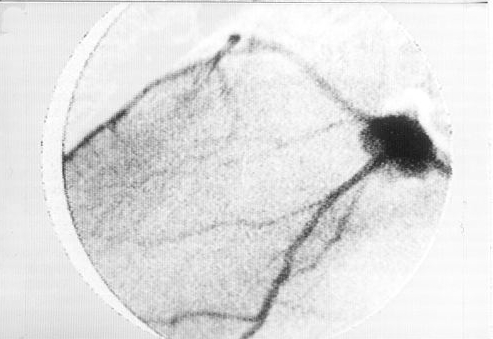
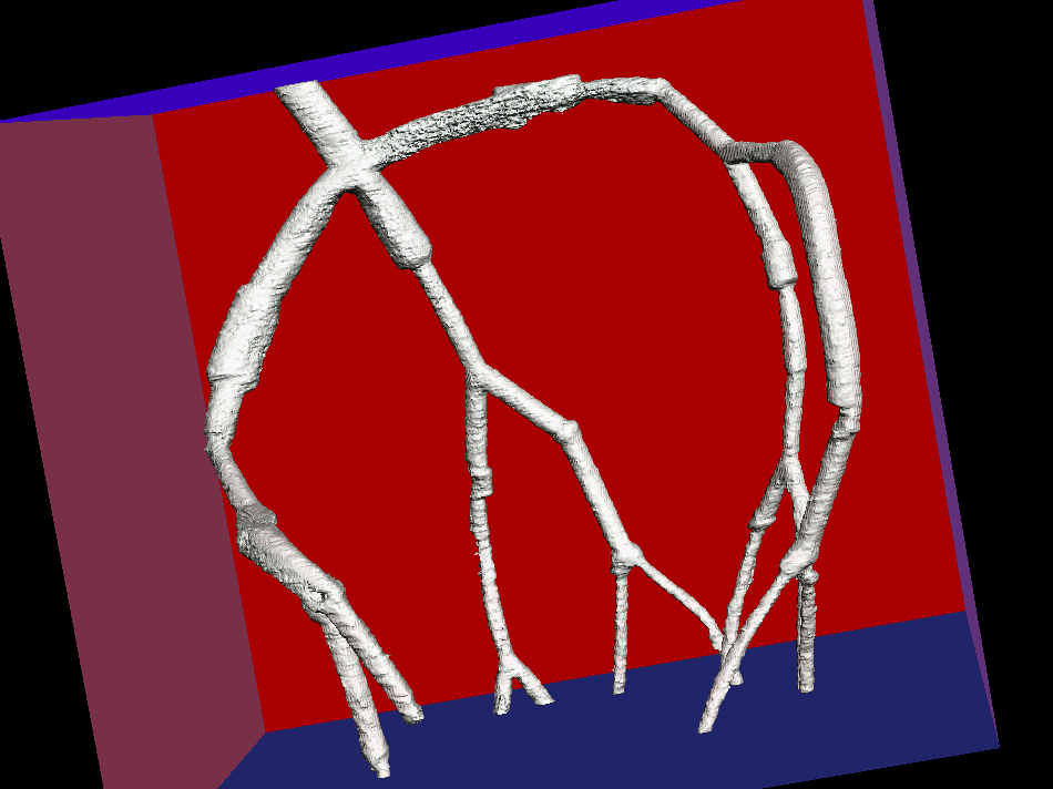
Canine myocardial contrast dye perfusion, pre-systole with coronary vessels.
Canine myocardial contrast dye perfusion at systole with coronary vessels.
Canine myocardial contrast dye perfusion, pre-systole.
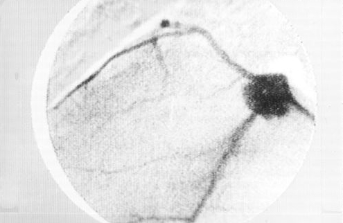
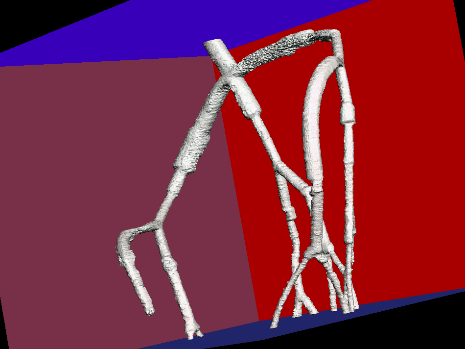
Coronary vessels removed, reconstructed with High-Efficiency CT from 3 bi-plane projections
Canine myocardial contrast dye perfusion at systole;
Coronary vessels removed, reconstructed with High-Efficiency CT from 3 bi-plane projections
Plastic Model
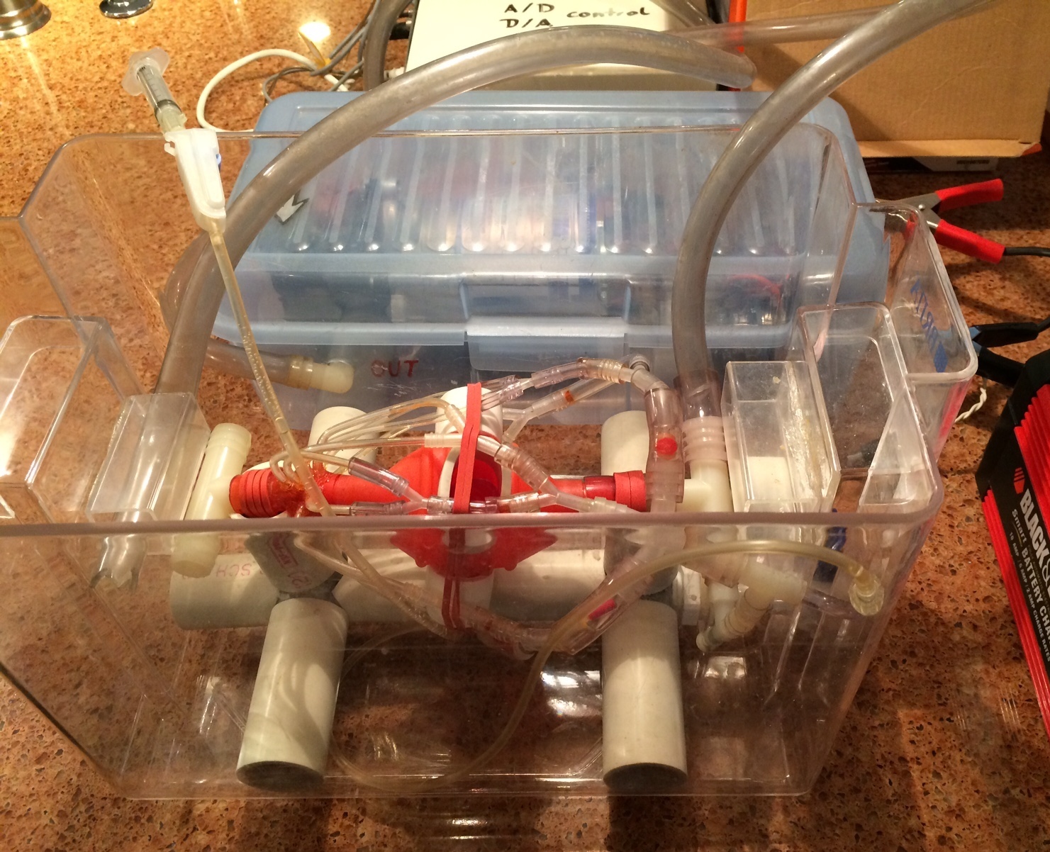
Physical plastic model with hydraulic pump to simulate coronary arteries of a beating heart, data acquisition and control system in the background, power supply for the hydraulic beat-pump to the right. Observe the red plastic stenoses inserted in the plastic tubes and the diameter changes of connected plastic tubes.
The model operates in the water-filled plastic container.
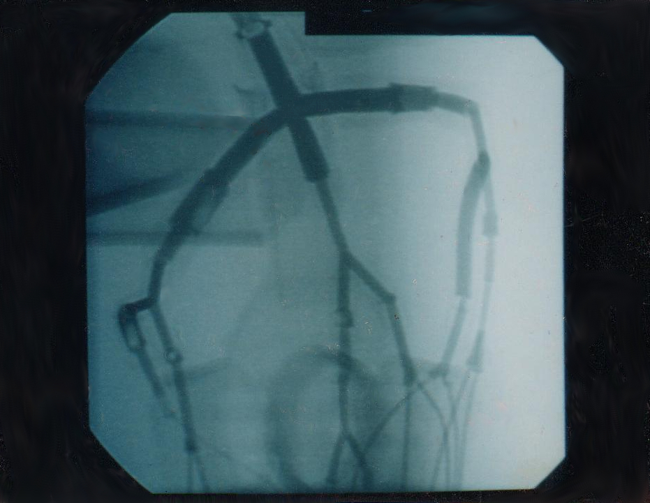
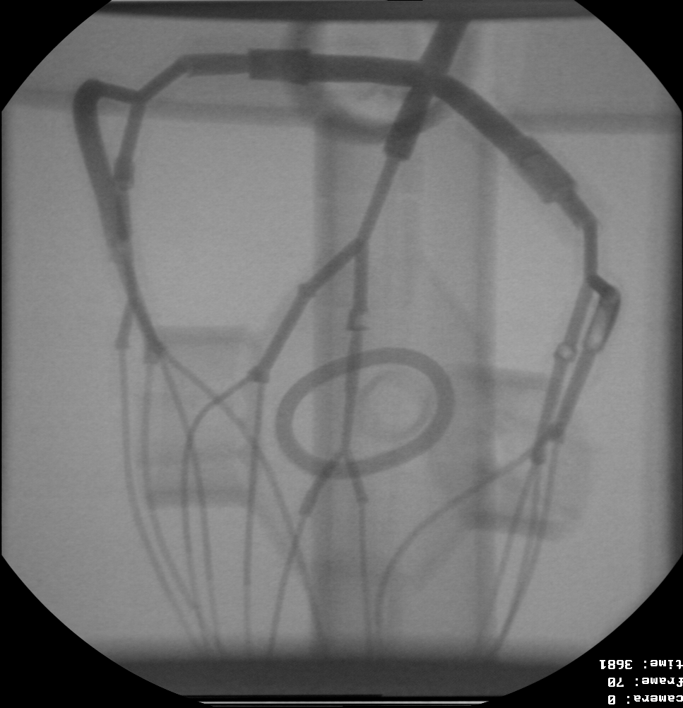
X-ray image of Hydraulic Model during contrast dye injection showing stenosis and changes in tube diameter. Raw digital image, recorded at 30 frames / second.
Front view of hydraulic model during contrast dye injection, logarithmic density correction (upright).
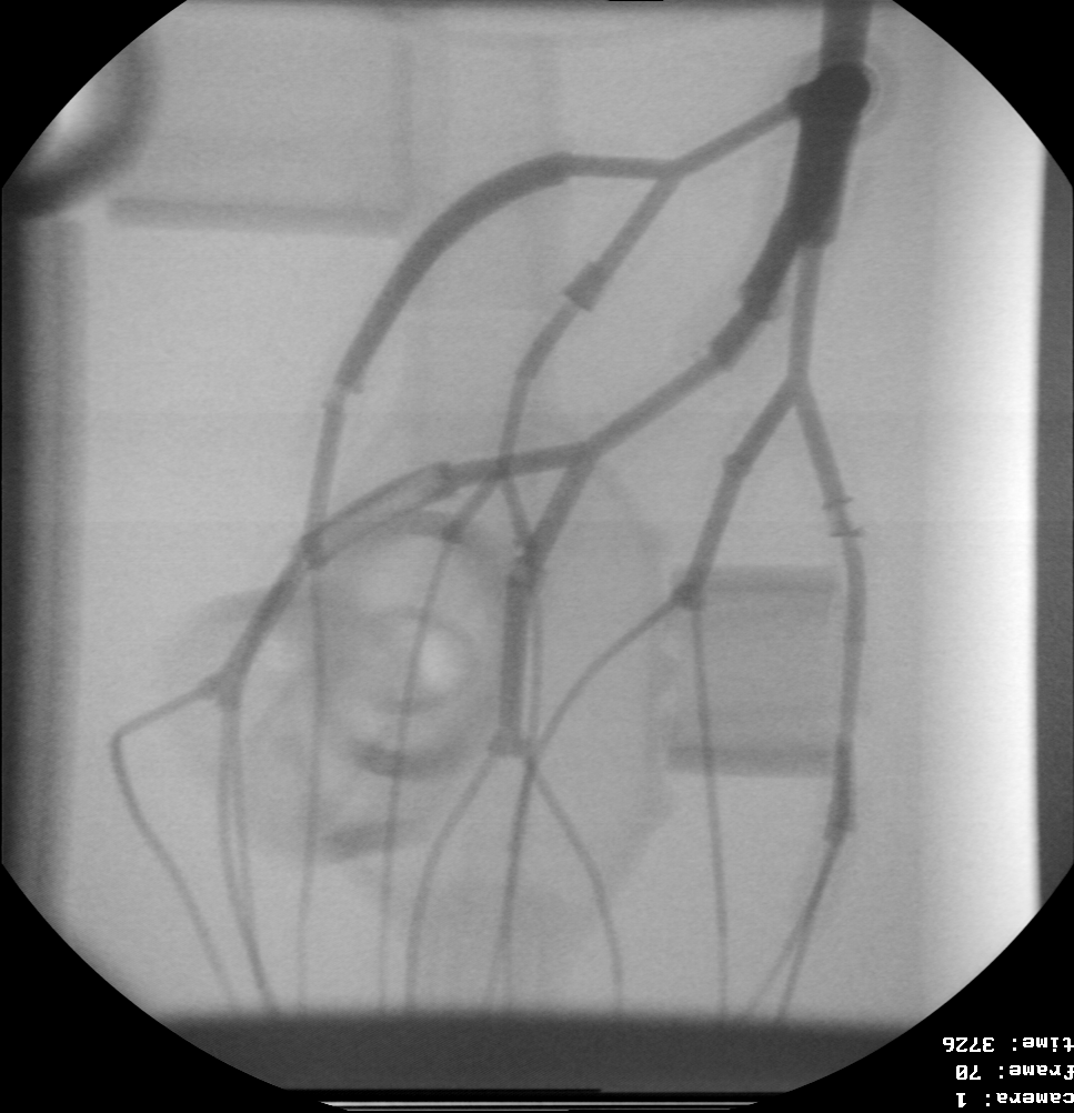
Side view of hydraulic model during contrast dye injection, logarithmic density correction (upright).
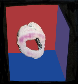
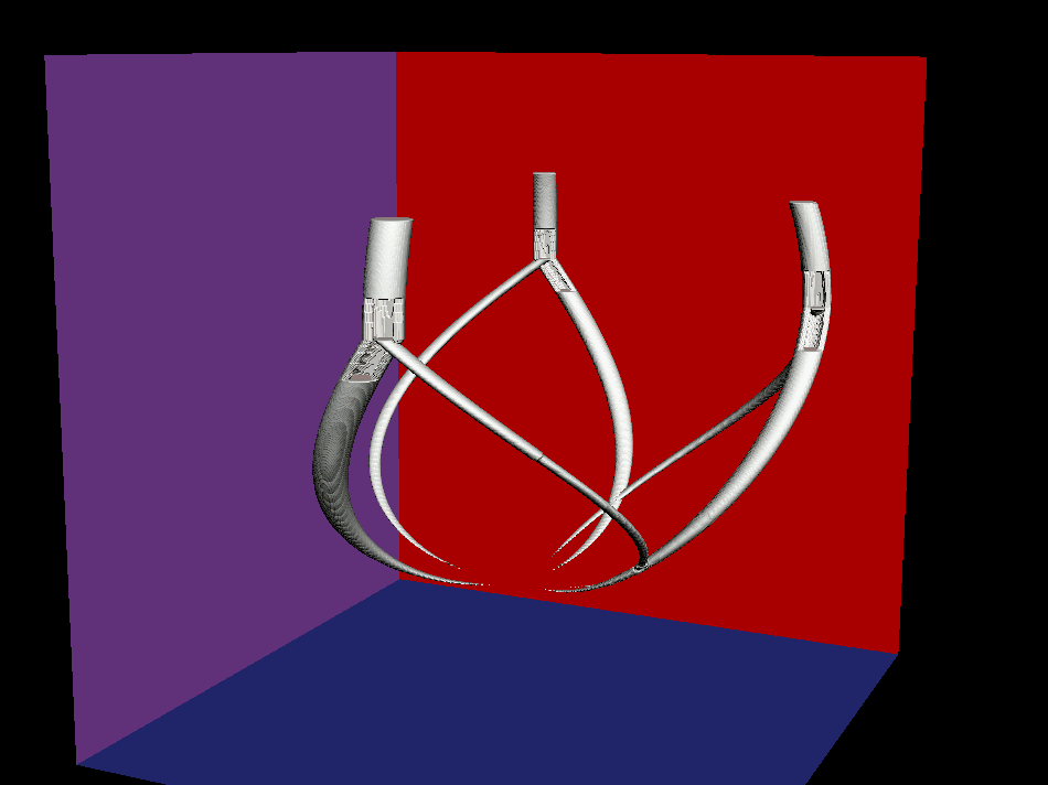
Perspective view of the reconstructed beating coronary artery model.
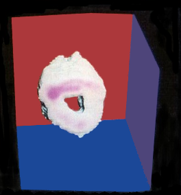
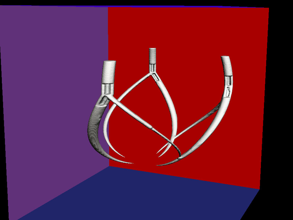
Perspective side-view of the reconstructed beating coronary artery model.
Numerical: Coronary Simulation
Numerical object simulation with three branches representing a coronary tree and three stenoses at branch-points.
Reconstruction of simulated branches and stenoses from 6 cone-beam projections.
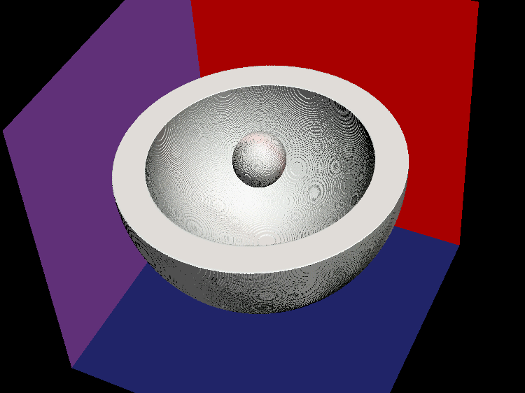
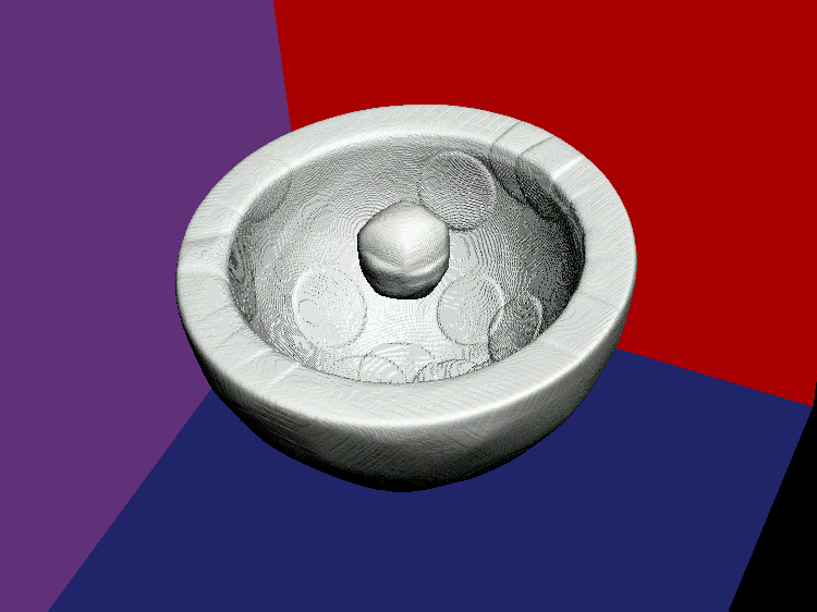
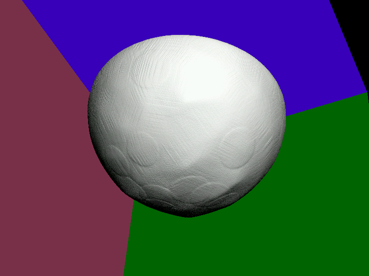
Numerical: Semisphere Simulation
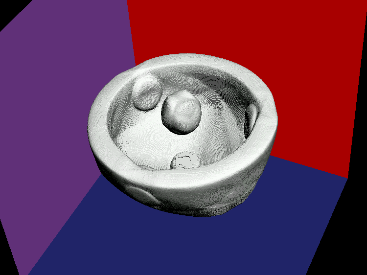
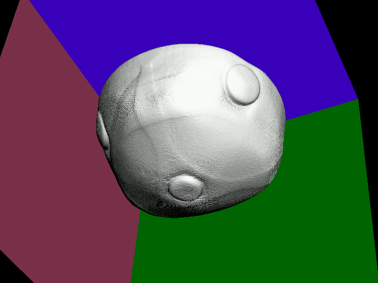
HIGH efficiency CT, startup iterations. Automated alignment w/o use of manually labeled beats.
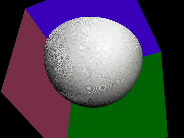
Numerical semisphere, bottom view.
Reconstruction from 4 cone-beam projections, top-view, 1 iteration.
Reconstruction from 4 cone-beam projections, bottom-view, 1 iteration.
Reconstruction from 18 cone-beam projections, top-view, 1 iteration.
Reconstruction from 18 cone-beam projections, bottom-view, 1 iteration.
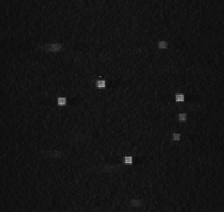
Numerical: Noise test / NIBIB
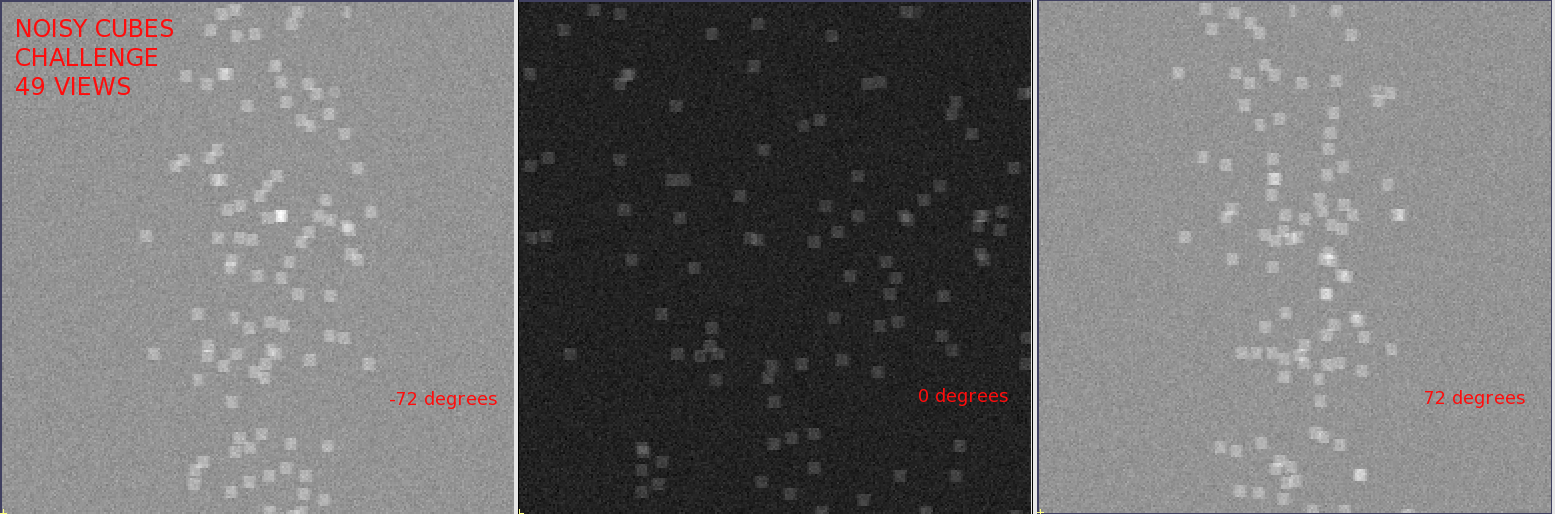
Three input projections of sparse 49 noisy cube projections, range from -72 to +72 degrees.
Provided by NIBIB of NIH
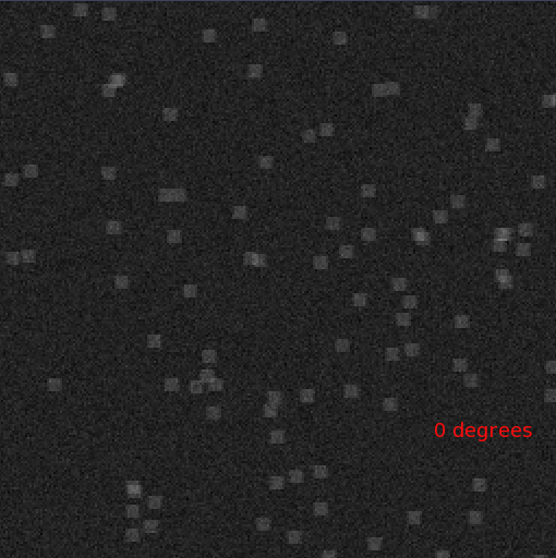
Magnified 0-degree projection of input data.
Magnified, central slice of HECT reconstruction from 49 projections.

Magnified, residuals at 0 degree projection of traditional maximum entropy (MENT) reconstruction after 15 iterations.
Provided by NIBIB of NIH for error comparison.

Magnified, corresponding residuals at 0 degree projection with High Efficiency Computed Tomography (HECT) after 1 full resolution reconstruction.
Team

Patent Information

Country
China
France
Germany
Japan
Taiwan
United Kingdom
USA
Patent No.
1555514
Patent Information
High Efficiency CT
US: 8,660,328, 8,660,330, 10,607,378, 10,789,743
IN: 334,717;
PCT: EP2310840
CN: 200980123522.8, ZL 201180035037.2
JP: JP5543448B2;
TW I555514;
Intl. pending: see WIPO/PCT;
JP2019535405A
Mammography: US: 11,123,032;
Relevant Sources – EP Smart Search, WIPO , USPTO, JPO